J105 3D Chemical Imager
|
|
|
|
|
|
|
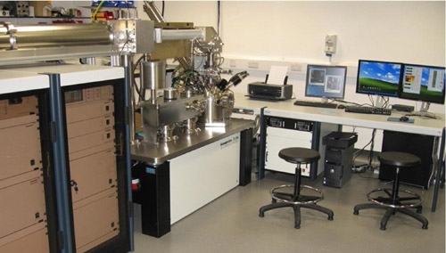
The J105 3D Chemical
Imager s a high performance SIMS system which uses DC
primary
cluster beams and a revolutionary time of flight analyser
to create a new paradigm
in organic analysis. The use of a DC primary
ion beam allows rapid imaging with
continuous data acquisition (i.e.
no separate etch cycle), with simultaneous high
spatial resolution
and high mass resolution. For imaging of organic samples,
the instrument is equipped with 40 kV C60 and gold cluster beams.
The analyser
is a dual-focussing combination of a shaped field buncher and a
non-linear reflectron.
Automated sample insertion,
a 1 micron precision 3-axis stage and camera assisted
sample positioning
allow simple and precise experimental setup. There is also
provision
for cold sample handling and fracture to facilitate analysis of
frozen hydrated
or freeze-dried samples. The J105 operates entirely
under computer control;
experiments can be routinely pre-programmed
and run automatically.
Features
• High mass resolution and high spatial resolution at
the same time.
• MS-MS
• Depth profiling
• 3D imaging
• Large area imaging
• Rapid, continuous acquisition
• Automated operation
• Advanced data viewing facility
The following data, provided
by the Surface Anaysis and Research Centre at The
University of
Manchester, illustrates the versatility and performance of the J105
Instrument in tissue and cell analysis |
|
|
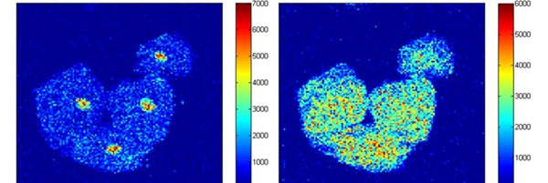
Imaging
of cheek cells
The
image to the left shows m/z 102.8 concentrated in the cell nuclei,
and mass 103.0
throughout the cell, demonstrating the need for high
mass resolution when imaging.
The field of view was approximately
170 x 150 microns.
|
|
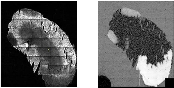
Imaging of unwashed
rat kidney section
|
|
By using a stage
scanning programme, a series of small area images can be tiled to
form a large area image. The data shown above were acquired by tiling
10 x 10
images, each 32 x 32 pixels, to form a total image with FOV
9 x 9 mm.
The images show the different distributions in the kidney
of m/z 184 (left) and
577 (right).
|
|
3D cell imaging
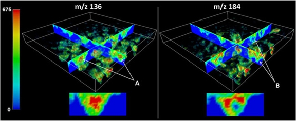
3D reconstruction
of image data from benign prostatic hyperplasia (BPH) cells.
Iso-surface
rendering shows the distribution of signal in 3D through the entire
sample
while orthogonal slices through the data set facilitate visualisation
of the chemical
distribution within cells.
|
|
|
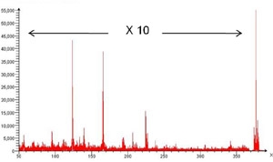
MS-MS of Haloperidol
When the MS-MS
mode is selected on the
J105, gas is introduced into a collision
cell
just before the first time focus, and an ion
gate on the time
focus selects the parent
mass of interest. The figure shows the
MS-MS spectrum of Haloperido;
showing parent peak and daughter
peaks
from collision induced dissociation. |
|
|
|
|